Comprehensive Guide To Breast Cancer Screening And Diagnostic Techniques
Role and Cost of Diagnostic Breast Cancer Procedures in the United States
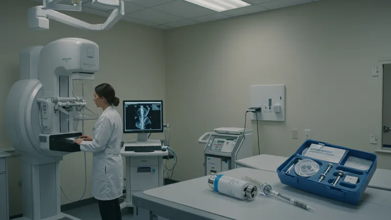
Diagnostic imaging, such as targeted mammography and stereotactic biopsy, is generally reserved for patients who have had abnormal screening results or present with specific symptoms. These advanced imaging strategies enable precise targeting of suspicious areas and enable radiologists to distinguish between benign and malignant changes.
Core needle biopsy remains a minimally invasive method to obtain tissue samples and is widely regarded as the diagnostic standard once imaging alone cannot definitively classify a lesion. The cost for this procedure can vary, but it is routinely performed in outpatient settings and is typically less costly than open surgical biopsies.
Stereotactic biopsy serves a critical role in sampling microcalcifications or small lesions that are difficult to localize on ultrasound. Using real-time X-ray guidance, this technique can be performed swiftly, often negating the need for more invasive surgery. While effective, the expense and access may fluctuate based on geographic region and insurance coverage.
When assessing distant disease spread or monitoring complex cases, PET scans offer unparalleled insights, although they are more commonly used in staging and surveillance. Their high cost limits frequent utilization; however, they are invaluable where detailed whole-body imaging is necessary to guide therapeutic decisions.